Foodborne Pathogens

Foodborne illnesses (commonly known as food poisoning) are often caused by eating food contaminated by bacteria and/or their toxins, parasites, viruses, chemicals, or other agents. Although the food supply in the United States is among the safest in the world, the federal government estimates that there are about 48 million cases of foodborne illness each year. This estimate equates to 1 in 6 Americans getting sick from contaminated food, resulting in about 128,000 hospitalizations and 3,000 deaths.

Food Pathogens occur when people eat or drink food or drink that is contaminated with pathogens, chemicals, or toxins. There are several factors that can contribute to the symptoms and severity of food poisoning, including a weakened immune system and age. When the FDA learns of an outbreak, the agency’s Coordinated Outbreak Response and Assessment Network (CORE) works closely with state and local partners and the Centers for Disease Control to identify the cause and prevent further illness. When necessary, FDA works with food manufacturers to facilitate voluntary recalls of potentially contaminated products; the agency also has mandatory recall authorities under the FDA’s Food Safety Modernization Act (FSMA).

Causes of food poisoning

Many different germs that cause illness can contaminate food, so there are many different foodborne infections (also called foodborne illness or food poisoning).

  • Researchers have identified more than 250 foodborne illnesses.
  • Most of them are infections caused by a variety of bacteria, viruses, and parasites.
  • Toxins and harmful chemicals can also contaminate food and cause foodborne illness.

Do I have food poisoning?

Common symptoms of foodborne illness are nausea, vomiting, stomach cramps, and diarrhoea. However, symptoms can differ between different types of foodborne illnesses. Symptoms can sometimes be severe, and some foodborne illnesses can even be life-threatening. Although anyone can get a foodborne illness, some people are more likely to develop it. Those groups include:

  • Older adults
  • Small children
  • People with weakened immune systems from medical conditions such as diabetes, liver disease, kidney disease, organ transplants, or HIV/AIDS, or from receiving chemotherapy or radiation therapy.
  • Pregnant women

Most people with foodborne illnesses get better without medical treatment, but people with severe symptoms should see their doctor.

Some Common Foodborne Germs

The top five germs that cause illness from foods eaten in the United States are:

  • norovirus
  • Salmonella
  • Clostridium perfringens
  • Campylobacter
  • Staphylococcus aureus (staphylococcus)

Some other germs don’t cause as much illness, but when they do, illnesses are more likely to lead to hospitalization. Those germs include:

  • Clostridium botulinum (botulism)
  • listeria
  • Escherichia coli (E.coli)
  • vibrio

Entry of enveloped viruses into host cells

Summary

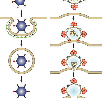
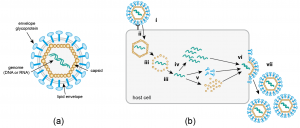
Viruses are intracellular parasites that hijack cellular machinery for their own replication. Therefore, a mandatory step in the virus life cycle is the delivery of the viral genome into the cell. Entry of enveloped viruses into cells (i.e., viruses with a lipid envelope) use a two-step procedure to release their genetic material into the cell: (i) they first bind to specific surface receptors on the target cell membrane and then, ( ii) fuse the viral and cellular membranes. This latter step may occur at the cell surface or after internalization of the viral particle by endocytosis or some other pathway (eg, macropinocytosis).

Remarkably, the virus-cell membrane fusion process follows essentially the same intermediate steps as other membrane fusions that occur, for example, in vesicle fusion at nerve synapses or cell-cell fusion in yeast mating. . Specialized viral proteins, fusogenic, promote fusion of the virus-cell membrane. Viral fusogenic undergo drastic structural rearrangements during fusion, releasing the energy needed to overcome the repulsive forces that prevent spontaneous fusion of the two membranes.

Keywords: class I fusion protein, class II fusion protein, class III fusion protein, enveloped virus, fusion pore, glycoprotein, membrane, membrane fusion intermediate, post-entry events, viral fusogen, virus entry

The Mechanical Properties Of The Influenza Virus Change According To Host Signals

Influenza virus passes through acidic endosomes, an essential step for shedding and infection. While residing in low pH conditions, protons pass through the viral M2 ion channel into the lumen of the virus and there separate the viral ribonucleoprotein (vRNP) cores from the M1 protein by inducing a conformational change in M1. This is consistent with the observation that the M1 protein has multiple functions in the virion and occurs in a ribbon or coiled structure.

Proton entry into the virion lumen then softens the viral envelope, most likely by dissociating vRNPs from the inner side of the envelope. Evidence for this notion was obtained by atomic force microscopy measurements with native virions and “bald” particles lacking viral glycoproteins. The latter result implies that low pH-induced changes in viral glycoproteins do not contribute significantly to envelope softening under low pH conditions.

Host Mechanics Control TheDeveloping Genome Of Adenovirus And Influenza Virus

Many viruses, including adenoviruses, infect postmitotic cells and replicate in the nucleus. Adenovirus deposits its linear DNA genome, but not the viral capsid, in the nucleus, as click-chemically tagged incoming virus genomes at single-molecule resolution recently demonstrated. The virus uses the molecular motor kinesin-1 to separate the genome from the capsid. This occurs on the cytoplasmic face of the nuclear pore complex (NPC), where the major capsid protein of the virus couples to the nucleoporin Nup214. The kinesin-associated 1,2 light chain binds to another virus capsid protein (protein IX), and the motor domain on the heavy chain is activated by binding to Nup358 of the filaments of the cytoplasmic pore complex.

Microtubules are located proximal to nuclear pore complexes because they have a binding site in the distal domain of Nup358. This quinary complex of viruses, two Nups and two motor components, then executes virus disruption and coincidentally part of the nuclear pore complex as well. The viral DNA (presumably in a complex with the viral protein VII) is then imported into the nucleus with the help of cellular transport factors, such as importins and transportin. However, the nuclear import process of viral DNA is not perfectly accurate and a variable fraction of the incoming viral DNA is misdelivered to the cytoplasm. This raises questions about innate immune recognition of cytosolic viral DNA and possible viral antagonism.

In addition to adenovirus, shedding of the influenza virus genome depends on cellular motor proteins, in this case, dynein and myosin. Under conditions of low pH in the endosome, the influenza virus alters the conformation of the glycoprotein hemagglutinin, leading to exposure of the hydrophobic fusion peptide and fusion of the viral membrane with the limiting endosomal membrane. Cytosolic RNPs dissociate from the membrane fusion site by a process that mimics the formation and removal of offenders.

This involves the unanchored ubiquitin-binding domain (ZnF-UBP) and dynein-binding domains of histone deacetylase 6 (HDAC6), as well as microtubule- and actin-based motors. Since the ionic milieu in endosomal compartments is subject to regulation (late but not early endosomes contain high concentrations of potassium ions and low concentrations of sodium ions, for example), additional signals acting on the virus can be anticipated. endosomal. In fact, the high concentrations of potassium ions together with the low pH in late endosomes prime the influenza A virus to separate vRNPs well before fusion.

Conclusions And Perspectives

This assay has attempted to integrate the physical properties of individual virus particles with mechanical or chemical signals from host cells. Physical and structural virology will continue to elucidate novel and exciting features of individual virus particles and show that they are important for infection, virus transmission, or vaccination against viral diseases. However, the full power and importance of these features are only manifested if the physical and structural features are integrated into the context of cellular or immunological mechanisms.

This approach considers the environment in which viruses have been selected in the course of evolution. Furthermore, it is important to note that viruses act as a swarm of particles and genetic elements, and a single virus particle rarely manages to infect a single cell. Rather, more often than not, virus particles cooperate or compete during infection. This notion gives rise to the emerging field of pathogen coinfection: for example, virus-virus and virus-bacteria coinfections.


Bacterial Transformation

Bacteria are commonly used as host cells to make copies of DNA in the laboratory because they are easy to grow in large numbers. Your cellular machinery naturally carries out DNA replication and protein synthesis.

Amazing bacteria

Bacteria are incredibly versatile organisms that have the unique ability to take in foreign DNA and replicate (or copy) it. This gives them an evolutionary advantage and helps them survive changes in their environment. For example, bacteria can acquire DNA that makes them resistant to antibiotics. The bacterial genome is contained in a single circular chromosome. This genetic material floats freely in the cell, unlike in eukaryotic organisms where the genetic material is enclosed within a nuclear membrane. Bacteria can sometimes contain smaller circles of DNA, called plasmids, that have a much smaller number of genes. Plasmids can be exchanged between bacteria in a process called conjugation.

Use of plasmids in the laboratory

Plasmids can be used as vectors to transport foreign DNA into a cell. Once inside the cell, the plasmid is copied by the host cell’s own DNA replication machinery. In the laboratory, plasmids are specifically designed for bacteria to copy the DNA they contain.

Essential elements of the plasmid

Laboratory-engineered plasmids contain a small number of genes that aid transformation. These include:

  • An origin of replication. This is the specific sequence of nucleotides where DNA replication begins.
  • A multiple cloning site. This site contains recognition sites for specific restriction enzymes. These restriction enzymes can be used to “cut” the plasmid so that the foreign DNA can be “glued on” by ligation.
  • A resistance gene. This gene encodes a protein that bacteria need to survive in a particular growth medium, for example when a specific antibiotic is present.

The piece of DNA or gene of interest is cut from its original DNA source using a restriction enzyme and then glued into the plasmid by ligation. The plasmid containing the foreign DNA is now ready to be inserted into bacteria. This process is called transformation.

Bacterial transformation

Before the bacterial transformation, the bacteria are treated with a chemical called calcium chloride, which causes water to enter the cells and swell them. These swollen bacteria are known as competent bacteria. Plasmid DNA (containing the foreign DNA) is then mixed with the competent bacteria and the solution is heated. Plasmid DNA enters the bacterium through tiny pores created in the cell membranes. Once in the host cell, the plasmid DNA is copied many times by the bacteria’s own DNA replication machinery.

How to know if it worked?

After transformation, the bacteria are grown on a nutrient-rich food called agar. Only bacteria that contain a plasmid with antibiotic resistance will grow in the presence of antibiotics. For example, if bacteria are grown on agar containing the antibiotic ampicillin, only bacteria that have been transformed with a plasmid containing the ampicillin resistance gene will survive. The transformed bacteria can then be grown in large numbers. The DNA of interest, or the protein encoded by the DNA, can be isolated and purified.

When is transformation used?

Bacterial transformation is used:

  • To make multiple copies of DNA is called DNA cloning.
  • To make large amounts of specific human proteins, for example, human insulin, which can be used to treat people with type I diabetes.
  • Genetically modify a bacterium or other cell.